Circulating Tumor Cells
By: Iman Ali, Galaxy International School

Introduction
Metastasis, the process by which cancer spreads, represents one of the most perilous aspects of the disease. Coined by Jean Claude Recamier in 1829, "metastasis" serves as the defining characteristic of a malignant tumor (Talmadge & Fidler, 2010). Circulating tumor cells (CTCs), also referred to as circulating cancer cells, play a pivotal role in this phenomenon. These are cells that have detached from the primary tumor and entered the circulatory or lymphatic systems. CTCs emerge as intermediaries within a complex multistep process, encompassing the transformation of primary tumor cells into tumor-initiating cells (TICs) via the epithelial-mesenchymal transition (EMT). These TICs subsequently become circulating tumor cells through intravasation into the bloodstream, and upon their arrival at distant sites, they establish themselves as disseminated tumor cells. This entire process leads to metastasis via the mesenchymal-epithelial transition (MET) (Agnoletto & Volinia, 2022).
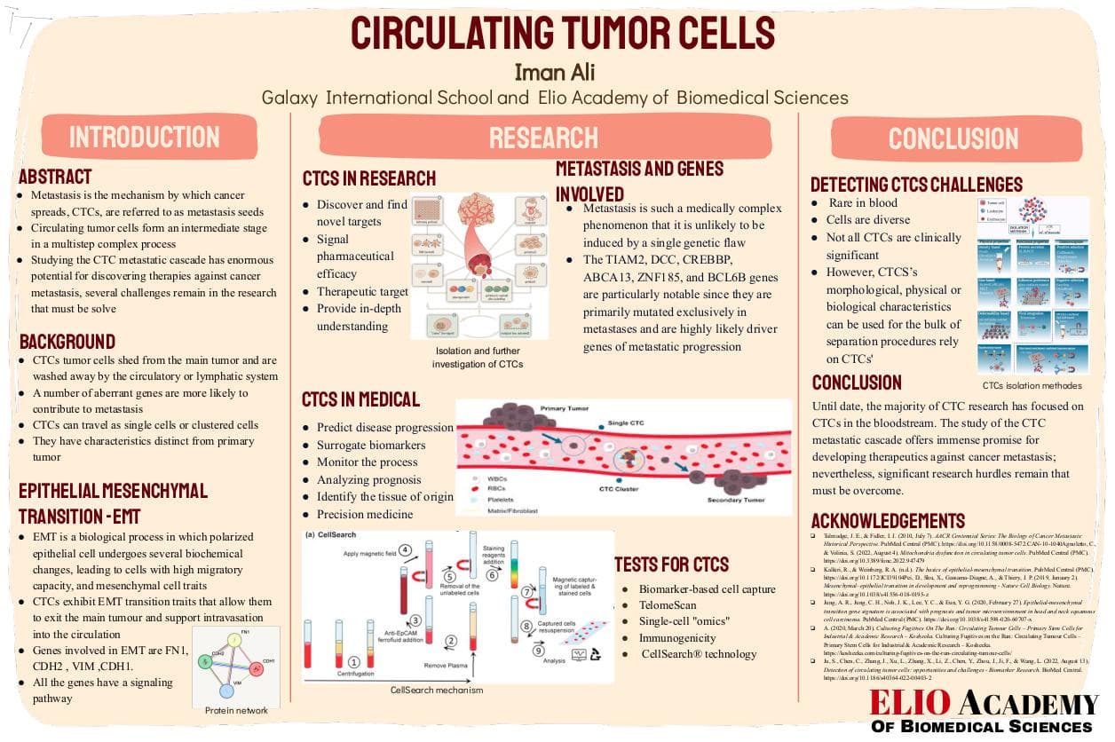
Upon reaching and settling within distant organs, disseminated tumor cells and CTCs collectively become known as metastasis seeds (Redirecting, n.d.). CTCs can traverse in two distinct forms: as solitary cells or as clusters, as illustrated in Figure 3 (2020). Remarkably, CTCs have been identified in the majority of tumor patients (Agnoletto & Volinia, 2022). Nevertheless, most existing CTC research has centered on the CTCs present within the bloodstream. A comprehensive study of the CTC metastatic cascade carries tremendous potential for unveiling novel therapies targeting cancer metastasis. However, numerous challenges persist within this field of research, requiring resolution (Ju et al., 2022).

The process of metastasis (Source)
Biology of CTCs and genes involved
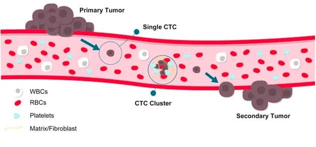
CTCs exhibit distinct characteristics compared to the primary tumor from which they originate. This distinction arises despite their derivation from the primary tumor. Epithelial-mesenchymal transition (EMT) is a biological process whereby polarized epithelial cells undergo several biochemical changes, resulting in cells with heightened migratory capacity and acquiring mesenchymal traits. Conversely, mesenchymal-epithelial transition (MET) is the reverse process of EMT (Kalluri & Weinberg, n.d.), (Pei et al., 2019). Critical genes participating in epithelial-mesenchymal transition encompass FN1 (Fibronectin 1), CDH2 (N-cadherin), VIM (vimentin), and CDH1 (E-cadherin) (Jung et al., 2020). Vimentin, classified as intermediate filaments of class III, is found in non-epithelial cells, particularly mesenchymal cells. Fibronectin 1 (FN1) contributes to cell adhesion, motility, opsonization, wound healing, and cell shape preservation. CDH2 (N-cadherin) is involved in neuronal recognition mechanisms and acts as a regulator of neural stem cell quiescence, Figure 3-- facilitating homotypic cell-cell adhesion. CDH1 (E-cadherin), in cell-cell connections, predominantly interacts homophilically, assisting in the sorting of heterogeneous cell types. All these genes involved in epithelial-mesenchymal transition exhibit interconnectedness, as depicted in Figures 2 and 3. CTCs manifest traits of EMT transition, facilitating their detachment from the primary tumor and supporting intravasation into circulation. They assemble in clusters to bolster their metastatic potential and harbor stemness characteristics that enhance their metastatic initiation ability (Lin et al., 2021).

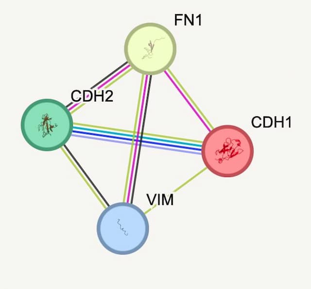
Figure 1 -Protein network (Source)

Figure 2 -Protein network edges (Source)
The intricacies of metastasis are such that a solitary genetic anomaly is unlikely to trigger it (Metastasis | Causes, Symptoms & Treatment, n.d.). Rather, a multitude of aberrant genes are more likely to collectively contribute to metastasis (Metastasis | Causes, Symptoms & Treatment, n.d.). Numerous studies have unveiled the complexity of abnormal gene interactions associated with metastasis. For instance, in a study of breast cancer patients with brain metastasis, defects in 17 genes were linked to this phenomenon (Metastasis | Causes, Symptoms & Treatment, n.d.). Of particular significance are genes such as TIAM2, DCC, CREBBP, ABCA13, ZNF185, and BCL6B, which exhibit exclusive mutations in metastases and are strong candidates as driver genes for metastatic progression (Krøigård et al., 2018). The BCL6B gene, also known as a transcription repressor located on chromosome 17, regulates gene expression, inflammatory responses, and type 2 immune responses (BCL6B BCL6B Transcription Repressor [Homo Sapiens (Human)] - Gene - NCBI, 2018). BCL6B methylation has been correlated with tumor growth, angiogenesis, metastasis, and invasion (Gu et al., 2018). Genes associated with metastasis have been categorized into three types based on their involvement across different stages of the metastatic process: metastasis initiation genes, metastasis advancement genes, and metastasis virulence genes (Krøigård et al., 2018).
CTCs Utilization in the medical field
CTCs\' Potential in Early Cancer Diagnosis (Lawrence et al., 2023): CTCs offer promise for early cancer diagnosis.
Predicting Disease Progression and Survival: Numerous studies spanning the last decade suggest that CTCs could serve as predictive markers for disease progression and survival, applicable to individuals with both metastatic and potentially early-stage cancers (Plaks et al., n.d.).
Surrogate Biomarkers: CTCs function as surrogate biomarkers across various solid cancer types, including breast cancer, melanoma, liver cancer, gastric cancer, pancreatic cancer, prostate cancer, and lung cancer (Circulating Tumor Cells: Biology and Clinical Significance - PubMed, 2021). Surrogate biomarkers gauge patient well-being, functionality, or survival and are designed to forecast treatment outcomes (Katz, n.d.).
Monitoring Cancer Progression (Lawrence et al., 2023): CTCs facilitate minimally invasive monitoring of cancer progression.
Analyzing Prognosis: CTCs assist in analyzing prognosis and tumor burden, even post-surgery (Nikanjam et al., 2022).
Identifying Tissue of Origin: CTCs are shed from diverse regions within tumors, exhibiting noticeable gene expression variations between primary tumors and CTCs, as well as within the CTC population. It may be feasible to identify the tissue of origin of CTCs using expression profiling to detect organ-specific metastatic signals (Plaks et al., n.d.).
Empowering Precision Medicine: Customized treatments are crucial, and CTCs offer insights into tumor RNA, DNA, and protein. CTCs and primary tumors often demonstrate comparable responses and resistances to medications, making them potential tools for individualized medication testing (Circulating Tumor Cells in Precision Medicine: Challenges and Opportunities - PubMed, 2022).
CTCs utilization in research field
Discovering Novel targets: CTCs analysis also has potential to discover and find new targets for therapeutic intervention (Krebs et al., n.d.).
Signaling Pharmaceutical Efficacy: CTCs have the potential to signal pharmaceutical efficacy even in the absence of visible metastases, and give insights into drug resistance processes.
Emerging Therapeutic Targets: They may also become a therapeutic target using single cell omics illustrated in figure 4. This might aid in the localization of small metastatic lesions and in guiding further diagnostic and treatment techniques (Plaks et al., n.d.).
Deepening understanding: The study of CTC functional and molecular properties may provide in-depth understanding about very deadly tumor disorders (Ju et al., 2022).

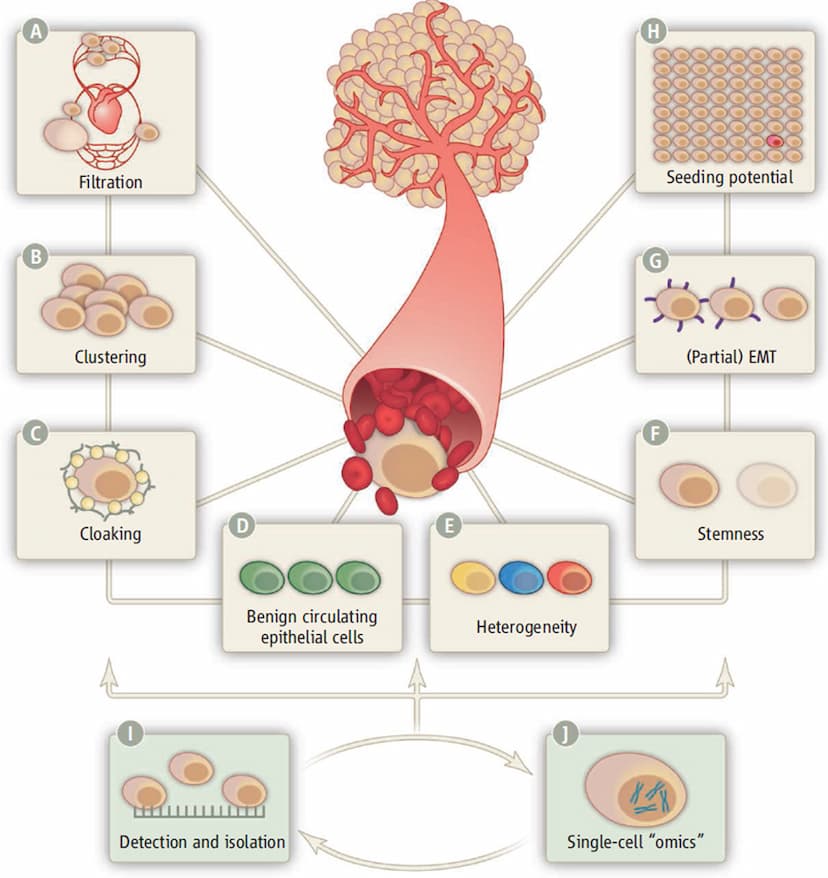
Figure 4 -- Isolation and further investigation of CTCs (Source)
Challenges with detecting CTCs in the blood and the separation strategies
Challenges and Considerations in CTC Analysis
Rarity in Blood:Immune system activity contributes to the swift demise of most CTCs within circulation, occurring within a short span of time after their release from tumors (Agashe & Kurzrock, 2020). Mechanisms like shear stress, anoikis, oxidative stress, as well as cytokine and growth factor shortages further contribute to their rapid elimination (Ju et al., 2022). As a result, the number of CTCs in the bloodstream is remarkably low, with only approximately 1-100 cells and 106-108 red blood cells per milliliter of blood (Circulating Tumor Cells: Biology and Clinical Significance - PubMed, 2021), making their detection challenging.
Cell Diversity:The diversity among cancer cells leads to significant variations in the expression of surface markers across different CTC groups. Consequently, distinguishing various types of CTCs based on the same criteria becomes a challenge (Circulating Tumor Cells: Biology and Clinical Significance - PubMed, 2021).
Clinical Significance of CTCs:Not all CTCs hold therapeutic relevance. While current research has identified viable circulating epithelial cells in patients with benign inflammatory colon diseases, they have not been found in healthy volunteers. Additionally, CTCs have been detected in the blood of a pancreatic cancer mouse model prior to the formation of a primary tumor. This makes it challenging for existing CTC assays to accurately differentiate between cancer cells, noncancerous tumor components, and benign cells (Plaks et al., n.d.).
Overcoming Technological BarriersScientists are leveraging technology to overcome challenges in CTC analysis. Due to the scarcity of CTCs within peripheral blood, isolating them from blood cell contamination is a crucial step for future research. However, the low frequency of CTCs and their inherent variability make high-precision detection time-consuming. Separation methods primarily rely on CTCs\' morphological, physical, or biological characteristics (Fig.6). Drawbacks such as low cell recovery, purity, and vitality hinder the widespread use of CTCs in both laboratory and clinical settings (Ju et al., 2022).

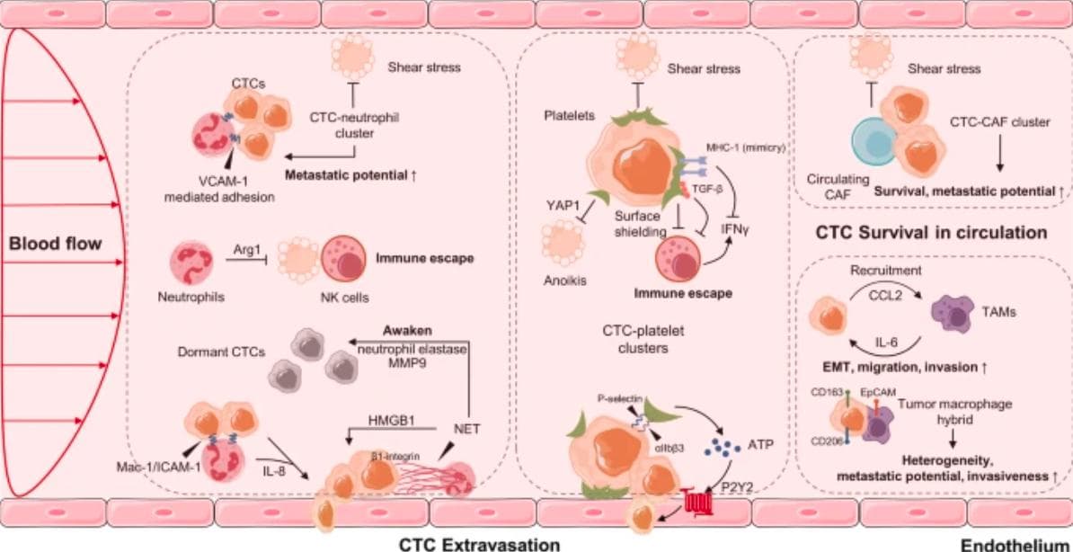
Fig 5: CTCs in The interaction of CTCs with neutrophils, platelets, CAFs, and a in the blood microenvironment. TAMs are tumor-associated macrophages, while CAFs are cancer-associated fibroblasts. (Source)
Specific Assays available for CTCs
Biomarker-based cell capture: CTCs were extracted using biomarker-based cell capture and enrichment based on biophysical and other properties (Nikanjam et al., 2022).
TelomeScan: Telomere length has frequently been utilized to estimate the future longevity of cells. TelomeScan identifies live CTCs in human peripheral blood using a telomerase-specific replication selective adenovirus. (Ju et al., 2022)

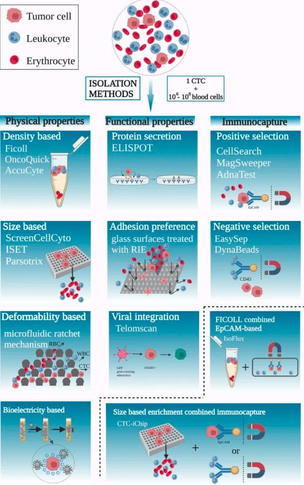
Figure 6 -- CTCs isolation methods (Source)
Single-cell \"omics\": The next frontier in CTC research is characterisation using single-cell \"omics\" methods that are constantly evolving. This will ultimately determine the clinical value of CTCs as biomarkers and therapeutic targets (Plaks et al., n.d.).
Immunogenicity: One of the most often used methods for separating CTCs is CTC enrichment by immunogenicity. Using particular biomarkers expressed on the cell surface, cells are captured and attached to a device surface or magnetic material. However, because CTCs express a variety of surface markers, there is no one, universal CTC antigen (Nikanjam et al., 2022).
CellSearch technology:
Another method, licenced by Janssen Diagnostics, is the CellSearch CTC technology, which is utilised for CTC identification and has been approved by the FDA to help in the monitoring of patients with metastatic breast, prostate, and colorectal cancers. Following centrifugation, the CellSearch® technology combines ferrofluid nanoparticles functionalized with an EpCAM antibody to facilitate magnetic separation of EpCAM+ cells from solid blood components. The cells are subsequently immunostained to confirm the presence of CK 8, 18, 19, and DAPI expression, as well as the absence of CD45 expression (Circulating Tumor Cell Technologies, 2016). Figure 7 depicts the procedure.
In a single center, observational study 132 patients were analyzed using cell search technology. As conclusion, the pleural CELLSEARCH test may be a beneficial supplement to standard cytology in terms of determining the diagnosis of malignant effusions. Another advantage is that it is relatively cheap and easily available (Schwed Lustgarten et al., n.d.).

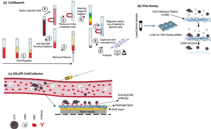
Figure 7: CellSearch mechanism for CTCs detection (Source)
Future directions of research
The Anticipated Impact of Malignant Cancer Testing
In the coming years, malignant cancer tests are expected to become accessible to a large population and will be routinely administered to enable early diagnosis. This early detection is vital as it allows for prompt treatment during the initial stages of cancer. CTCs, as highlighted in the report, serve various purposes and offer a means to monitor, track, and identify metastatic cancer. Given that metastatic disease accounts for over 90% of cancer-related deaths (Krebs et al.), CTCs offer a potential avenue to reduce this statistic, offering hope for improved outcomes. A critical avenue lies in the development of sensitive and highly specific devices that leverage the amassed knowledge. These devices should possess the capability to detect various types of circulating tumor cells---a formidable challenge due to the diversity among these cells.
Another approach involves identifying shared traits among all CTC types while differentiating them from other cell types. This intricate device, when realized, will be able to isolate CTCs from the vast sea of cells, enabling the identification of tumor type and origin. Once developed, the device will be used not only for detection but also for gaining insights into tumor mutations and origins through single-cell omics. A profound comprehension of the diseases is pivotal to advancing effective cures. CTCs are poised to offer a new dimension in monitoring treatment progress. If treatment efficacy is suboptimal, the test results will indicate this to doctors, enabling swift adjustments to treatment plans, potentially leading to more effective outcomes. The integration of these opportunities into medical practice can substantially elevate the treatment of malignant tumors, resulting in the potential to save numerous lives.
The upcoming era of malignant cancer testing, spearheaded by CTC analysis, holds immense promise for revolutionizing cancer diagnosis, treatment, and patient outcomes.


Impact Statement

My name is Iman, and I am a senior at Galaxy International School. I felt connected to the healthcare aspect more than anything else, as in it I saw a purpose and a versatile vocation. My appetite for scientific knowledge and my ability to work patiently and methodically are aspects of my character that drew me to study biomedical science specifically. To get a head start on my aim to become a biomedical scientist, I applied to the Elio Academy of Biomedical Science to study from professionals in the field. Every session left me wanting to learn more. As I delved into the world of biomedical science, I was also given the opportunity to work on my own research project. During our initial lectures, we learned about circulating tumour cells. It piqued my interest and drew me in to explore it. I thoroughly enjoyed writing a report on it and would like to thank ELIO Academy for providing me with this amazing opportunity.
Student Reflection
References
Talmadge, J. E., & Fidler, I. J. (2010, July 7). AACR Centennial Series: The Biology of Cancer Metastasis: Historical Perspective. PubMed Central (PMC). [https://doi.org/10.1158/0008-5472.CAN-101040
Agnoletto, C., & Volinia, S. (2022, August 4). Mitochondria dysfunction in circulating tumor cells. PubMed Central (PMC). [https://doi.org/10.3389/fonc.2022947479
Kalluri, R., & Weinberg, R. A. (n.d.). The basics of epithelial-mesenchymal transition. PubMed Central (PMC). [https://doi.org/10.1172JCI39104
Pei, D., Shu, X., Gassama-Diagne, A., & Thiery, J. P. (2019, January 2). Mesenchymal--epithelial transition in development and reprogramming - Nature Cell Biology. Nature. [https://doi.org/10.1038/s41556-018-0195z
Jung, A. R., Jung, C. H., Noh, J. K., Lee, Y. C., & Eun, Y. G. (2020, February 27). Epithelial-mesenchymal transition gene signature is associated with prognosis and tumor microenvironment in head and neck squamous cell carcinoma. PubMed Central (PMC). [https://doi.org/10.1038/s41598-020-60707x
A. (2020, March 20). Culturing Fugitives On The Run: Circulating Tumour Cells -- Primary Stem Cells for Industrial & Academic Research -- Kosheeka. Culturing Fugitives on the Run: Circulating Tumour Cells -- Primary Stem Cells for Industrial & Academic Research -- Kosheeka. [https://kosheeka.com/culturing-fugitives-on-the-run-circulating-tumour-cell/
Ju, S., Chen, C., Zhang, J., Xu, L., Zhang, X., Li, Z., Chen, Y., Zhou, J., Ji, F., & Wang, L. (2022, August 13). Detection of circulating tumor cells: opportunities and challenges - Biomarker Research. BioMed Central. [https://doi.org/10.1186/s40364-022-004032
Lin, D., Shen, L., Luo, M., Zhang, K., Li, J., Yang, Q., Zhu, F., Zhou, D., Zheng, S., Chen, Y., & Zhou, J. (2021, November 22). Circulating tumor cells: biology and clinical significance - Signal Transduction and Targeted Therapy. Nature. [https://doi.org/10.1038/s41392-021-008178
Metastasis | Causes, Symptoms & Treatment. (n.d.). Encyclopedia Britannica. [https://www.britannica.com/sciencemetastasis
Krøigård, A. B., Larsen, M. J., Lænkholm, A. V., Knoop, A. S., Jensen, J. D., Bak, M., Mollenhauer, J., Thomassen, M., & Kruse, T. A. (2018, January 2). Identification of metastasis driver genes by massive parallel sequencing of successive steps of breast cancer progression. PubMed Central (PMC). [https://doi.org/10.1371/journal.pone0189887
BCL6B BCL6B transcription repressor [Homo sapiens (human)] - Gene - NCBI. (2018, April 4). BCL6B BCL6B Transcription Repressor [Homo Sapiens (Human)] - Gene - NCBI. [https://www.ncbi.nlm.nih.gov/gene255877
Gu, Y., Li, A., Sun, H., Li, X., Zha, H., Zhao, J., Xie, J., Zeng, Z., & Zhou, L. (2018, February 1). BCL6B suppresses proliferation and migration of colorectal carcinoma cells through inhibition of the PI3K/AKT signaling pathway. PubMed Central (PMC). [https://doi.org/10.3892/ijmm.20183451
Lawrence, R., Watters, M., Davies, C. R., Pantel, K., & Lu, Y. J. (2023, June 2). Circulating tumour cells for early detection of clinically relevant cancer - Nature Reviews Clinical Oncology. Nature. [https://doi.org/10.1038/s41571-023-00781y
Plaks, V., Koopman, C. D., & Werb, Z. (n.d.). Circulating Tumor Cells. PubMed Central (PMC). [https://doi.org/10.1126/science1235226
Circulating tumor cells: biology and clinical significance - PubMed. (2021, November 22). PubMed. [https://doi.org/10.1038/s41392-021-008178
Katz, R. (n.d.). Biomarkers and Surrogate Markers: An FDA Perspective. PubMed Central (PMC). [https://doi.org/10.1602/neurorx.1.2189
Nikanjam, M., Kato, S., & Kurzrock, R. (2022, September 12). Liquid biopsy: current technology and clinical applications. PubMed Central (PMC). [https://doi.org/10.1186/s13045-022-01351y
Circulating tumor cells in precision medicine: challenges and opportunities - PubMed. (2022, May 1). PubMed. [https://doi.org/10.1016/j.tips.2022.02005
Krebs, M. G., Hou, J. M., Ward, T. H., Blackhall, F. H., & Dive, C. (n.d.). Circulating tumour cells: their utility in cancer management and predicting outcomes. PubMed Central (PMC). [https://doi.org/10.11771758834010378414
Agashe, R., & Kurzrock, R. (2020, August 21). Circulating Tumor Cells: From the Laboratory to the Cancer Clinic. PubMed Central (PMC). [https://doi.org/10.3390cancers12092361
Circulating tumor cell technologies. (2016, January 28). Circulating Tumor Cell Technologies - ScienceDirect. [https://doi.org/10.1016/j.molonc.2016.01007
Schwed Lustgarten, D. E., Thompson, J., Yu, G., Vachani, A., Vaidya, B., Rao, C., Connelly, M., Udine, M., Tan, K. S., Heitjan, D. F., & Albelda, S. (n.d.). Use of Circulating Tumor Cell Technology (CELLSEARCH) for theDiagnosis of Malignant Pleural Effusions. PubMed Central (PMC). [https://doi.org/10.1513/AnnalsATS.201303068OC
Redirecting. (n.d.). Redirecting. [https://doi.org/10.1016/j.celrep.2022111298
By: Iman Ali
The opinions expressed here are the views of the writer and do not necessarily reflect the views and opinions of Elio Academy.