Burkitt Lymphoma
Understanding its Relation to the c-Myc Gene

Introduction to Burkitt Lymphoma
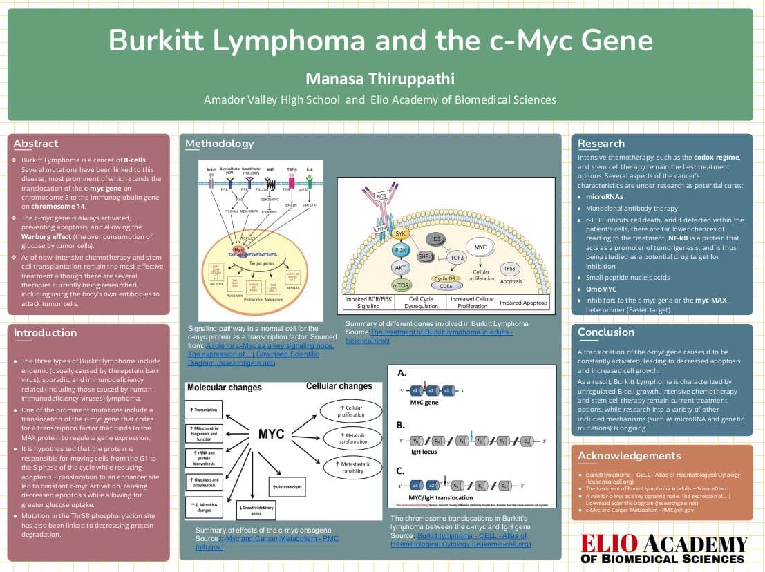
Burkitt Lymphoma represents a rare yet highly aggressive form of Non-Hodgkin's cancer originating in the immune system. This cancer type triggers uncontrolled growth of B-cells and is marked by symptoms such as abdominal pain, nausea, vomiting, loss of appetite, weakness, and swollen lymph nodes. Tumor lysis, wherein tumor cells release their contents into the bloodstream, often leads to organ damage and loss of muscle control. An intriguing characteristic of Burkitt Lymphoma is the presence of irregular macrophages that have ingested cancer cells, alongside cancer cells displaying large vacuoles. This unique appearance creates a "starry sky" pattern in lymph nodes. The disease encompasses three distinct types: endemic (often linked to the Epstein-Barr virus), sporadic, and immunodeficiency-related lymphomas (including those associated with human immunodeficiency viruses).

Summary of different genes involved in Burkitt Lymphoma ([Source](https://www.sciencedirect.com/science/article/pii/S0006497121002615?via%3Dihub))
Testing
Diagnosing Burkitt Lymphoma typically involves a lymph node biopsy, followed by computed tomographic (CT) imaging of the chest, abdomen, and pelvis, chest X-rays, PET or gallium scans, bone marrow biopsies, spinal fluid examinations, and blood tests to assess kidney and liver function. Additionally, testing for HIV disease and measuring lactate dehydrogenase levels in the blood are undertaken.
Genetic Basis of Burkitt Lymphoma and c-myc gene
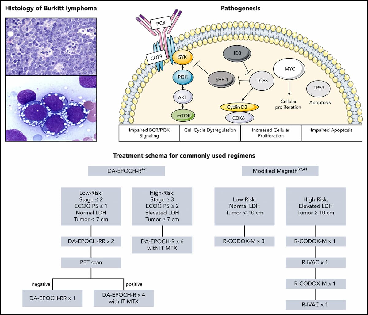
While various genes play a role in Burkitt Lymphoma, alterations in the c-myc gene have been widely observed as a potential contributor to unregulated cell division. The c-myc proto-oncogene, located on chromosome 8, encodes the c-myc transcription factor. This protein heterodimerizes with the MAX transcription factor to bind to DNA at the E box consensus sequence, a critical site for gene regulation.
C-Myc and regulation
The promoter region of the c-myc gene contains multiple binding sites for transcription factors and ribonucleoproteins, including Oct, SREBP, MPB-1, and others. Retention of RNA polymerase at the start of exon 1 impedes mRNA transcription from other exons. The c-myc protein's brief half-life and mRNA stability contribute to its regulatory mechanisms.
c-myc Protein and Cell Cycle Function:

Signaling pathway in a normal cell for the c-myc protein as a transcription factor. ([Source](https://www.researchgate.net/figure/Fig-1-A-role-for-c-Myc-as-a-key-signaling-node-The-expression-of-intracellular-c-Myc_fig1_267739031))
The c-myc protein, comprised of 439 amino acids, assumes a basic helix-loop-helix structure. Upon binding with MAX, it forms an alpha helix shape. The protein's short half-life and phosphorylation for proteasomal degradation are key aspects. C-myc expression inhibits apoptosis and mutations are associated with reduced dependence on growth factors, fostering uncontrollable division. While the first exon is noncoding, the second and third exons encode the c-myc protein. The protein acts as a transcription factor for several genes while repressing cyclin D1. Thr58 phosphorylation also regulates c-myc protein turnover.
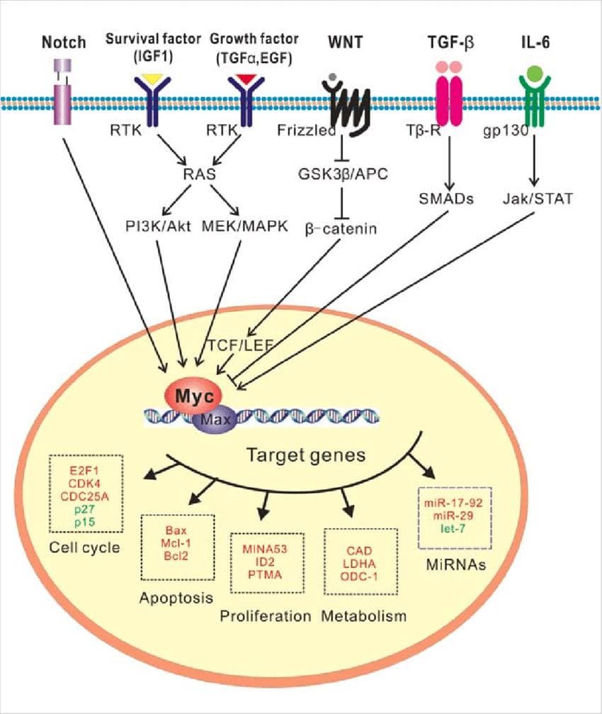
Mutation of the c-myc gene

Summary of effects of the c-myc oncogene ([Source](https://www.ncbi.nlm.nih.gov/pmc/articles/PMC3505847/))

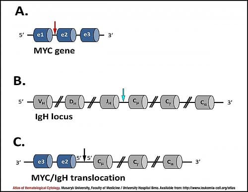
The chromosome translocations in Burkitt's lymphoma between the c-myc and IgH gene ([Source](https://www.leukemia-cell.org/atlas/index.php?pg=images--mature-b-cell-neoplasms--burkitt-lymphoma#1))
Treatment
- The primary approach for treating Burkitt Lymphoma involves undergoing intensive chemotherapy to eradicate malignant cells.
- Some of the commonly employed therapeutic regimens encompass dose-adjusted EPOCH, Cancer and Leukemia Group B (CALGB) 10002, CODOX-M, and HyperCVAD. These regimens incorporate an array of distinct drugs.
- Notably, the widely utilized drug, Rituximab, has demonstrated substantial advancements in patient outcomes, showcasing an enhanced effectiveness of approximately 10 to 15%.
- Nonetheless, among older patients, irrespective of the chosen treatment plan, there is a noticeable elevation in treatment-related mortality rates as compared to their younger counterparts.
- Subsequent to chemotherapy, the implementation of stem cell transplantation has yielded commendable progress, boasting an overall survival rate of approximately 83%. This finding stems from a study conducted between 1985 and 2007 by the Center for International Blood and Marrow Transplant Research.
- Conversely, patients who experience relapse often exhibit limited responsiveness to various treatment approaches.

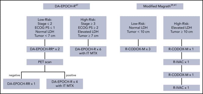
Common treatment options for BL patients ([Source](https://www.sciencedirect.com/science/article/pii/S0006497121002615?via%3Dihub#cesec40))
Ongoing research for therapy
Recent research has been focused on studying how the cancer cells in Burkitt Lymphoma evade apoptosis, specifically with the help of the c-FLIP and nuclear factor kappa B (NF-kB) pathway. c-FLIP inhibits cell death, and if detected within the patient's cells, there are far lower chances of reacting to the treatment. (NF-kB is a protein that acts as a promoter of tumorigenesis, and is thus being studied as a potential drug target for inhibition. However, this protein also acts as a transcription factor for Fas signaling pathway that induces apoptosis, and can therefore impair the body's biological response.
Another area of research is the presence of microRNAs and their role in tumor development). The differences in their expression in cancerous and healthy cells have potential to be further investigated and perhaps later act as a target for therapy.
Monoclonal therapy is another source of possible treatment, with increasing success being observed by the agent Rituximab. In such cases, an engineered monoclonal antibody marks cancerous cells for destruction by the immune system, destroying the cell membrane, blocking cell growth, or causing self-destruction.
Some epigenetic modifiers are also under study, such as depsipeptide (a histone deacetylase inhibitor) which is shown to kill cancerous cells. Finally, small peptide nucleic acids (chemicals that can be bound to DNA very tightly) are currently being researched in mice to reduce c-myc production through binding to complementary regulatory sequences in the intron. By invading the double helix of the DNA strands, it can bind to the required regulator of the c-myc gene and silence it.
Future directions of research
Currently, clinical trials are investigating bromo and extra terminal bromodomain inhibitors to down-regulate the c-myc gene. However, due to uncertainties regarding the gene's complete functions and potential unintended consequences of reducing its impact, these inhibitors cannot yet be employed in humans. Challenges also arise from the gene's nuclear location, hindering the synthesis of drugs with appropriate properties for nucleus penetration. The intricate folding of the c-myc protein further complicates drug binding for degradation/silencing. Targeting the complex of c-myc protein and max transcription factor, as seen with sAJM589, has shown promise in reducing myc expression and enhancing protein degradation. OmoMYC, a small protein, is being researched as an alternative, binding to MAX to counteract myc protein effects and inhibit cell growth. However, being a peptide, OmoMYC may face degradation by cellular machinery. While intensive chemotherapy remains the most effective Burkitt Lymphoma treatment, rising toxicity and treatment-related mortality have spurred the exploration of alternative therapies. For relapsing patients, combining chemotherapy with stem cell transplantation stands as the current optimal solution. Future research might explore a combination of inhibitors targeting both c-myc and other genes implicated in Burkitt Lymphoma, offering potential avenues for advancement.
Conclusion
Burkitt Lymphoma, a B-cell cancer, is marked by various gene mutations, notably the c-myc gene translocation to an immunoglobulin gene. This alteration leads to persistent c-myc gene activation, triggering reduced apoptosis and heightened cell division. Additionally, these gene mutations support the Warburg effect, causing elevated glucose consumption by tumors. High-dose chemotherapy is particularly effective, especially in pediatric patients. However, relapsed cases have shown better outcomes with chemotherapy combined with stem cell transplantation, while older patients often experience more adverse reactions and treatment toxicity. Ongoing research focuses on diverse avenues for future therapies, including microRNA targeting, monoclonal antibody therapy, small peptide nucleic acids, and down-regulating the c-myc gene. A deeper understanding of the cancer's mechanisms will help streamline potential treatments and drive the development of future therapeutic approaches.


Impact Statement

Hi! My name is Manasa Thiruppathi, and I'm a senior at Amador Valley High School. I've always been passionate about the curious enigma called the human body, and all the intricate mysteries behind its functioning. So when the opportunity came this summer to understand the basic controller of it all, genetics, I jumped at the chance. At the ELIO Academy of Biomedical Sciences, I explored the basic intricacies of genomics and the role it plays in a variety of afflictions, including cancer and immunodeficiencies, which culminated in a research paper (The Relation of the c-Myc Gene to Burkitt Lymphoma), using bioinformatic tools like GWAS and dSNP. In particular, I attempted to explain one of the many reasons for the cancer causing uncontrollable growth of B-cells; that is, the c-Myc gene that produces proteins related to the cell cycle. In the future, I hope to further explore the many hidden conundrums that biology holds.
Student Reflection
References
"Burkitt Lymphoma." Atlas of Haematological Cytology, www.leukemia-cell.org/atlas/index.php?pg=images--mature-b-cell-neoplasms--burkitt-lymphoma#5
"Burkitt Lymphoma." Cleveland Clinic, https://my.clevelandclinic.org/health/diseases/22777-burkitt-lymphoma
Burkitt lymphoma pathogenesis and therapeutic targets from structural and functional genomics. Pubmed, https://pubmed.ncbi.nlm.nih.gov/22885699/ .
"Burkitt's Lymphoma." Healthline, www.healthline.com/health/burkitts-lymphoma.
Crombie, Jennifer, and Ann LaCasce. The treatment of Burkitt lymphoma in adults. Pubmed, https://pubmed.ncbi.nlm.nih.gov/33171490/
Crown, John, et al. MYC as a target for cancer treatment. Pubmed, https://pubmed.ncbi.nlm.nih.gov/33524794/
Cryptic MYC insertions in Burkitt lymphoma: New data and a review of the literature. Pubmed, https://pubmed.ncbi.nlm.nih.gov/35167621/
Emison, E., et al. Function of the c-Myc oncogenic transcription factor. Pubmed, https://pubmed.ncbi.nlm.nih.gov/10579912/
Hamlyn, P. H., et al. Altered nucleotide sequences of a translocated c-myc gene in Burkitt lymphoma. Pubmed, https://pubmed.ncbi.nlm.nih.gov/6419122/
Henehan, Gary T., and Barry J. Ryan. Overview of approaches to preventing and avoiding proteolysis during expression and purification of proteins. Pubmed, https://pubmed.ncbi.nlm.nih.gov/23377852/
Johnston, J. M., and W. L. Carroll. c-myc hypermutation in Burkitt's lymphoma. Pubmed, https://pubmed.ncbi.nlm.nih.gov/1297477/
Lynch, David T., and Brittney S. Graham. Burkitt Lymphoma. Pubmed, https://pubmed.ncbi.nlm.nih.gov/30844175/
Mushtaq, Muhammad, et al. Different Mechanisms of Regulation of the Warburg Effect in Lymphoblastoid and Burkitt Lymphoma Cells. Pubmed, https://pubmed.ncbi.nlm.nih.gov/26312753/
Nguyen, Lynh, et al. The Role of c-MYC in B-Cell Lymphomas: Diagnostic and Molecular Aspects. Pubmed, https://pubmed.ncbi.nlm.nih.gov/28379189/
Targeted Disruption of Myc-Max Oncoprotein Complex by a Small Molecule. Pubmed, https://pubmed.ncbi.nlm.nih.gov/28976731/
Thompson, E. B. The many roles of c-Myc in apoptosis. Pubmed, https://pubmed.ncbi.nlm.nih.gov/9558477/
"Understanding Lymphoma Burkitt Lymphoma." Lymphoma Research Foundation, https://lymphoma.org/wp-content/uploads/2022/07/LRF_Burkitt_Lymphoma_Fact_Sheet.pdf
Von der Lehr, N., et al. c-Myc hot spot mutations in lymphomas result in inefficient ubiquitination and decreased proteasome-mediated turnover.
Yustein, Jason T., and Chi V. Dang. Biology and treatment of Burkitt's lymphoma. Pubmed, https://pubmed.ncbi.nlm.nih.gov/17534164/ .
Zayac, Adam S., and Adam J. Olszewski. Burkitt lymphoma: bridging the gap between advances in molecular biology and therapy. Pubmed, https://pubmed.ncbi.nlm.nih.gov/32255708/ .
_By: Manasa Thiruppathi_
The opinions expressed here are the views of the writer and do not necessarily reflect the views and opinions of Elio Academy.