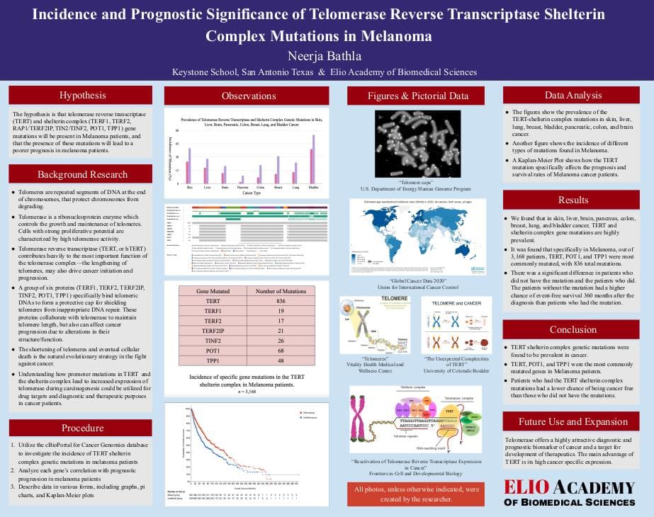
TERT Shelterin Complex & Carcinogenesis
By: Neerja Bathla

Neerja Bathla
Keystone SchoolMy project comprehensively investigates the role of TERT and telomere biology in Melanoma by examining the intricate relationship between telomerase, and shelterin complex genes in human genomic data. Through ERP, I enjoyed learning how to conduct scientific statistical analysis, use different biomedical databases, and enhance my knowledge and passion of biomedical research and the steps to conducting trials and creating new treatments and vaccines, among many other things. It has also helped me to develop an award winning science fair project and has inspired me for future projects. Elio Academy has been a part of my life for the past two years, through engaging, accommodating, and helpful instructors, who encouraged thinking and asking questions, I had an amazing opportunity to develop my identities as a student and researcher.
Short Report

Cancer Incidence
Cancer is the leading cause of death globally. There were an estimated 20 million new cases of cancer and 10 million deaths from cancer in 2020. An overall increase in life expectancy with evolution extends the period over which oncogenes act on cells. This increases the risk of cancer development. Cancer is caused by the accumulation of genetic mutations in cells. Cancer primarily happens when normal cells due to mutations acquire the ability to replicate indefinitely. This cellular immortality is the hallmark of cancer. The shortening of telomeres and eventual cellular death is the natural evolutionary strategy in the fight against cancer. The basis of cancer is the unlimited proliferation of cells, which in a lot of cases is achieved by the activation of telomerase leading to telomere stability and cellular immortality.

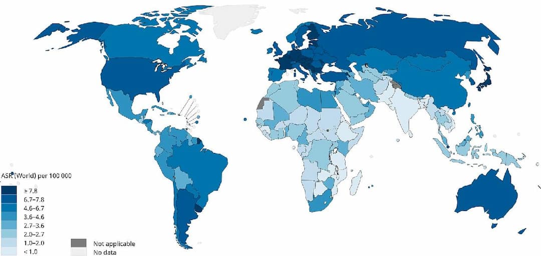
Incidence of Cancer Worldwide
Epidemiology of Melanoma (December 2021)
Melanoma accounts for 1.7% of global cancer diagnoses. Melanoma is the fifth most common cancer in the United States of America, growing over 320% in the nation since 1975. It has risen in developed countries with a majorly fair-skinned population. US mortality has fallen almost 30% over the past decade with the approval of ten new targeted drugs and immunotherapy agents since 2011. Mutations in the signaling protein BRAF are present in half of Melanoma cases, and are targeted with BRAF/MEK inhibitor combinations. Although the overall 5-year survival has risen to 93.3% in the US, survival for stage IV disease remains only 29.8%.
Melanoma is most common in white, older men, with an average age of diagnosis of 65. Outdoor UV exposure without protection is the main risk factor, although indoor tanning beds, immunosuppression, family history and rare congenital diseases, moles, and obesity contribute to the disease. Primary prevention initiatives in Australia implemented since 1988, such as education on sun-protection, have increased sun-screen usage and curbed melanoma incidence, which peaked in Australia in 2005. In the US, melanoma incidence is not projected to peak until 2022–2026.
Fewer than 40% of Americans report practicing adequate protection (sun avoidance from 10 a.m.–4 p.m. and regular application of broad-spectrum sunscreen with an SPF > 30). A 2-4-fold return on investment is predicted for a US sun-protection education initiative. Lesion-directed skin screening programs, especially for those at risk, have also cost-efficiently reduced melanoma mortality.

Gene Mutations
Telomere History and Discovery
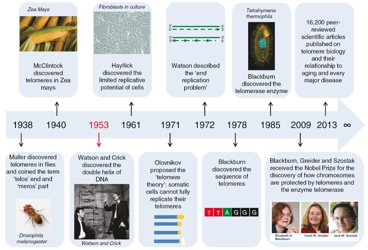
While conducting a radiation experiment on Drosophila chromosomes in 1938, Herman Muller found that X rays could induce chromosome rearrangements but not terminal deletions. This led him to believe in the special ability of terminal ends of chromosomes. He coined the term “telomere” for this region (derived from Greek words ‘telos’, meaning end, and ‘meros’, meaning part). In 1971, Russian biologist Alexey Olovnikov was the first to acknowledge the issue of telomere shortening, the end replication problem in chromosomes, predict the existence of the enzyme telomerase, and also came up with hypotheses associating telomere shortening with cellular senescence, or aging, and associating telomeres with carcinogenesis. Then, biologists Szostak, Blackburn, and Greider (2009) described that telomeres shortened 50–200 base pairs in each division until reaching the critical limit.

Telomere shortening has been assumed to act as a “mitotic clock” that limits the cell cycle number and critically short telomeres eventually trigger cellular senescence. In 2004, Liu et al. Corpus described that in somatic cells, telomerase remains inactive, but its activity can be found in germ cells and stem cells. In addition, reactivation of telomerase in somatic cells is one way to acquire uninhibited proliferation in cancer. Telomerase activity was detected in approximately 85% of malignant tumors including melanomas and cancers of the central nervous system, bladder, liver and thyroid.
Telomere Structure and Function
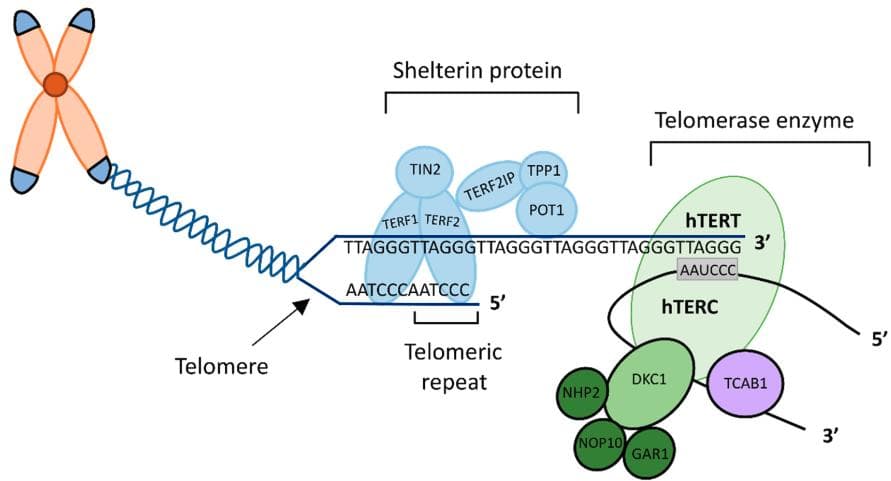
Telomeres are repeated segments of DNA found at the end of chromosomes. They protect chromosomes from degrading or breaking down. In normal cells, telomeres become progressively shorter as the cell divides. After a certain number of cell divisions, the telomeres become so short that they trigger the cell to stop dividing and die (apoptosis).Telomeres consist of thousands of repeats of the same short DNA sequence, which varies between organisms but is TTAGGG in humans and other mammals. Telomeres have two pivotal functions. On one side, telomeres are involved in the protection of chromosomal ends, as they prevent unwanted recombination and degradation. On the other side, telomeres play an important role during DNA replication as they prevent the loss of DNA. The three-dimensional folding of telomeres protects the free 3′-OH end of each DNA strand from being recognized as a double strand break (DSB).
Telomerase
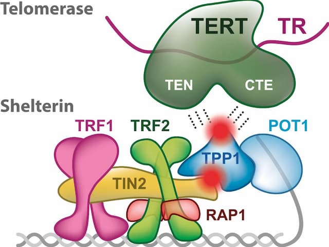
Telomerase is a ribonucleoprotein enzyme with reverse transcription activity. The DNA end-replication problem is solved by the action of telomerase. Telomerase contains an RNA template that synthesizes tandem repeats of telomeric DNA and compensates for the DNA shortening. Telomerase contains a reverse transcriptase (human telomerase reverse transcriptase, hTERT), an associated template RNA (telomerase RNA component, TERC) and accessory proteins.

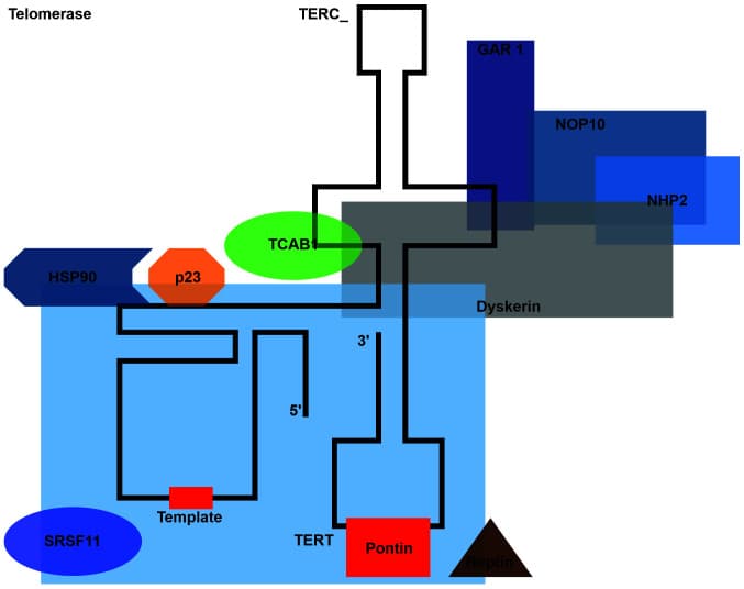
Telomerase structure Trybek, T., Kowalik, A., Góźdź, S., Kowalska, A. "Telomeres and telomerase in oncogenesis (Review)". Oncology Letters 20.2 (2020): 1015-1027.
Telomerase is a RNA-dependent DNA polymerase, meaning an enzyme that can make DNA using RNA as a template. It extends (adds nucleotides to) the overhanging strand of the telomere DNA using complementary RNA as a template. Telomerase is inactive in most somatic cells. However, hematopoietic stem cells, keratinocytes in the basal layer of the epidermis, uterine endometrial cells, germ cells and various tumors avoid telomere shortening by activation of telomerase. Studies have shown that inducing telomerase activity in primary human fibroblasts by retroviral gene transfer is sufficient to counteract telomere erosion and to prevent cells from entering senescence. The resulting maintenance of telomere length immortalizes most human cell types. Cells with strong proliferative potential are characterized by high telomerase activity.
The enzyme is active during embryonic development. Cancer cells are characterized by high telomerase activity, which enables cells to divide indefinitely. Telomerase is active in 85–95% of cancers. The exception is cancer cells possessing an active Alternative Lengthening of Telomeres (ALT) pathway. ALT, which is the ability of cancer cells to extend telomeres in the absence of telomerase, is based on homologous recombination using telomeric DNA as a matrix. ALT activation correlates with the presence of mutations in the genes encoding α-thalassemia/mental retardation X-linked chromatin remodeler and death domain associated protein in both tumors and cell lines . This process is observed in aggressive, difficult to treat tumors of mesenchymal origin, which account for approximately 5–15% of all cancers.

Telomerase Reverse Transcriptase (TERT)
Telomerase reverse transcriptase (abbreviated to TERT, or hTERT in humans) is a catalytic subunit of the enzyme telomerase, which, together with the telomerase RNA component (TERC), comprises the most important unit of the telomerase complex. The telomerase enzyme consists of two major components that work together. The component produced from the TERT gene is known as hTERT. The other component is produced from a gene called TERC and is known as hTR. The hTR component provides a template for creating the repeated sequence of DNA that telomerase adds to the ends of chromosomes. The hTERT component then adds the new DNA segment to chromosome ends. TERC is widely expressed in most cell types and even in telomerase-negative cells, whereas TERT expression is highly regulated, being absent or only present in low levels in somatic cells. The human TERT gene is located on chromosome 5p15.33. TERT induction and telomerase activation not only create unlimited cancer cell proliferation potential by stabilizing telomere length but also cause oncogenic effects independently of the telomere lengthening function.

Pavel Veverka, "Quantitative Biology of Human Shelterin and Telomerase: Searching for the Weakest Point." International Journal of Molecular Sciences, 28 June 2019, source
The telomere lengthening-independent functions of TERT, which significantly contribute to cancer initiation or progression, include its effects on mitochondrial and ubiquitin-proteasomal function, DNA damage repair, gene transcription, microRNA (miRNA) expression, RNA-dependent RNA polymerase activity, and epithelial-mesenchymal transition . These TERT activities physiologically affect the processes that ultimately lead to cell aging; however, they also drive cancer development by conferring survival, proliferation, motherhood, and invasive phenotypes. The TERT gene is located on the short arm of chromosome 5. The 433-bp genomic region located immediately upstream of the TERT core promoter region may bind to transcription factors or repressors. This region upstream of the TERT promoter is unmethylated in normal human cells, whereas it is methylated in malignant cells.
Shelterin Complex
As mentioned above, telomerase contains a highly conserved reverse transcriptase (human telomerase reverse transcriptase, hTERT), an associated template RNA (telomerase RNA component, TERC) and accessory proteins. A group of proteins specifically bind double-stranded and single-stranded telomeric DNAs to form a protective cap for shielding telomeres from inappropriate DNA repair. This group of proteins in mammals is called the shelterin complex. These proteins also collaborate with telomerase to maintain telomere length. The removal and mutation of these telomeric proteins activate DNA damage response pathways and trigger the degradation or fusion events of chromosomes. Shelterin is a six-protein complex - telomeric repeat-binding factor 1 (TRF1; also known as TERF1), telomeric repeat-binding factor 2 (TRF2; also known as TERF2), repressor and activator protein 1 (RAP1; also known as TERF2IP), TRF1-interacting nuclear protein 2 (TIN2; also known as TINF2), protection of telomeres 1 (POT1), and TPP1. TRF1 and TRF2 exist as homodimers and bind the double-stranded DNA regions of telomeres with high affinity and specificity. TRF1 negatively regulates telomere length and promotes the efficient replication of telomere DNA. TRF2 primarily protects telomeres from being recognized as double-stranded DNA breaks (DSB). RAP1 is recruited to telomeres through interaction with TRF2. RAP1 functions together with TRF2 to inhibit homologous recombination.
Telomere Biology and Carcinogenesis
One of the hallmarks of cancer is proliferation without restriction or immortalisation. One of the ways to attain this is through reactivation of telomerase in somatic cells. Telomerase activity has been consistently detected in 80–90% of malignant tumors. Understanding the molecular mechanism of how promoter mutations in TERT lead to increased expression of telomerase during carcinogenesis is important. As these alterations could potentially be exploited for diagnostic and therapeutic purposes in cancer patients.
TERT Mutations
Transcriptional regulation of the TERT gene occurs at many levels and is mediated by various positive and negative factors or signaling pathways .These factors control the TERT gene and ensure inhibition of TERT activity in most normal cells, as well as its expression at the right time and place in a small number of cell types such as activated lymphocytes or stem cells. This balance may be disturbed in malignant cells. A typical example is the Myc/Max/Mad1 protein. Endogenous expression of the cellular c-MYC oncogene may result in dissociation of the Mad1/Max repressor from the E-box complex, leading to de-repression of the TERT gene and telomerase activation. Epigenetic factors responsible for DNA methylation can also modulate TERT transcription. TERT promoter methylation is required for the expression of TERT and activation of telomerase in cancer cells. Some viruses may code for proteins that act as cofactors to stimulate TERT transcription as well. These include Epstein-Barr virus, cytomegalovirus, Kaposi sarcoma-associated herpesvirus, human papillomavirus, hepatitis B virus, hepatitis C virus, and human T-cell leukemia virus-1.
Shelterin Complex Mutations
Several mutations are reported in all three domains of RAP1 which are associated with cancer. Some of the critically important mutations are, p.M5I, p.D10H, p.Q191R, and p.R364X, found in the patient of familial melanoma, whereas mutations including p.A104P, p.R133Q are reported in chronic lymphocytic leukemia (CLL). POT1 was the first identified member of the shelterin complex which is mutated in cancer. Mutation in POT1 plays a critical role in telomeric DNA binding. POT1 mutations are likely causative of many diseases including CLL, familial melanoma, CMM, CP, cardiac angiosarcoma, and familial glioma. Among the six shelterin complex genes we analyzed, high TERF1 and TERF2 expressions were significantly associated with a poor prognosis (TERF1, HR 1.16 [95% CI: 1.04–1.3], p = 0.0062; TERF2, HR 1.16 [95% CI: 1.1–1.36], p = 0.056.

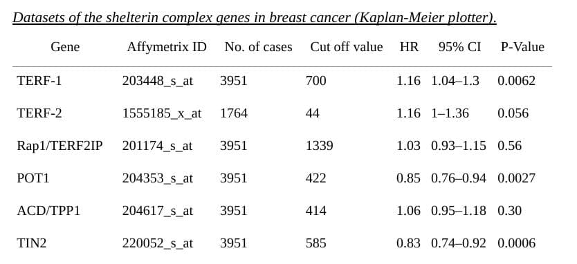
In addition, the mRNA expression levels of TPP1, and RAP1 were not correlated with RFS. The analysis identified that TERF1 and TERF2 may have prognostic potential in breast cancer. This is correlated with a recent study that shows that TERF-1 inhibition using combinatorial therapy can reduce stemness in glioblastoma xenograft models. In conclusion, the data analysis showed that overexpression of TERF1 and TERF2 mRNA is correlated to a poor prognosis for all BC patients and may be a useful tool for prognosis. The possible responsiveness to these shelterin complex genes expressed by breast cancer cells can be the consequence of activation of DNA damage and suppression of DDR pathways.
Non-telomere Related Functions of TERT
Several researchers found that TERT can act as a transcriptional regulator modulating the expression of genes in different pathways. These are involved in most physiological processes, including cell cycle, metabolism, differentiation, cell signaling and cell survival. Overall, the transcriptional abilities of TERT seem ascertained, not only because authors from different research groups observed these features, but also because mechanisms through which this action is exerted have been elucidated. Indeed, it has been shown that some genes are activated through the direct interaction between TERT and their promoters, for example, RB/E2F. Alternatively, other genes are triggered in an indirect manner, for example NF-kB and Wnt, in which TERT binds different proteins involved in their signaling cascade.
Studies from which Data was Obtained
- Acral Melanoma (TGEN, Genome Res 2017)
- Basal Cell Carcinoma (UNIGE, Nat Genet 2016)
- Cutaneous Squamous Cell Carcinoma (DFCI, Clin Cancer Res 2015)
- Cutaneous Squamous Cell Carcinoma (MD Anderson, Clin Cancer Res 2014)
- Cutaneous Squamous Cell Carcinoma (UCSF, NPJ Genom Med 2021)
- Desmoplastic Melanoma (Broad Institute, Nat Genet 2015)
- Melanoma (Broad/Dana Farber, Nature 2012)
- Melanoma (MSK, Clin Cancer Res 2021)
- Melanoma (MSK, NEJM 2014)
- Melanomas (TCGA, Cell 2015)
- Metastatic Melanoma (DFCI, Nature Medicine 2019)
- Metastatic Melanoma (DFCI, Science 2015)
- Metastatic Melanoma (MSK, JCO Precis Oncol 2017)
- Metastatic Melanoma (UCLA, Cell 2016)
- Skin Cutaneous Melanoma (Broad, Cell 2012)
- Skin Cutaneous Melanoma (TCGA, Firehose Legacy)
- Skin Cutaneous Melanoma (TCGA, PanCancer Atlas)
- Skin Cutaneous Melanoma (Yale, Nat Genet 2012)
- Skin Cutaneous Melanoma(Broad, Cancer Discov 2014)
Bibliography
Cancer stats https://www.paho.org/en/campaigns/world-cancer-day-2023-close-care-gap
Trybek, T., Kowalik, A., Góźdź, S., Kowalska, A."Telomeres and telomerase in oncogenesis (Review)". Oncology Letters 20.2 (2020): 1015-1027.
Mir SM, Samavarchi Tehrani S, Goodarzi G, Jamalpoor Z, Asadi J, Khelghati N, Qujeq D, Maniati M. Shelterin Complex at Telomeres: Implications in Ageing. Clin Interv Aging. 2020 Jun 3;15:827-839. doi: 10.2147/CIA.S256425. PMID: 32581523; PMCID: PMC7276337.
https://www.researchgate.net/publication/323523320_Telomere_biology_and_age-related_diseases#pf2
Yong Chen* The structural biology of the shelterin complex https://doi.org/10.1515/hsz-2018-0368 Received September 10, 2018; accepted October 15, 2018; previously published online October 20, 2018
Shelterin complex gene: Prognosis and therapeutic vulnerability in cancer; Vikas Kumar Bhari a, Durgesh Kumar b, Surendra Kumar c, Rajeev Mishra, Chen, Yong.
"The structural biology of the shelterin complex" Biological Chemistry, vol. 400, no. 4, 2019, pp. 457-466. https://doi.org/10.1515/hsz-2018-0368
Mir SM, Samavarchi Tehrani S, Goodarzi G, Jamalpoor Z, Asadi J, Khelghati N, Qujeq D, Maniati M. Shelterin Complex at Telomeres: Implications in Ageing. Clin Interv Aging. 2020 Jun 3;15:827-839. doi: 10.2147/CIA.S256425. PMID: 32581523; PMCID: PMC7276337.
Amir M, Khan P, Queen A, Dohare R, Alajmi MF, Hussain A, Islam A, Ahmad F, Hassan I. Structural Features of Nucleoprotein CST/Shelterin Complex Involved in the Telomere Maintenance and Its Association with Disease Mutations. Cells. 2020 Feb 4;9(2):359. doi: 10.3390/cells9020359 . PMID: 32033110; PMCID: PMC7072152.
By: Neerja Bathla
The opinions expressed here are the views of the writer and do not necessarily reflect the views and opinions of Elio Academy.


Full Report / White Paper


(Click to view the full report)
By: Neerja Bathla. The opinions expressed here are the views of the writer and do not necessarily reflect the views and opinions of Elio Academy.